At the office of Flossophy Dental, we combine experienced clinical judgment with modern imaging tools to deliver clearer diagnoses and more confident treatment plans. Cone-beam computed tomography (CBCT) is one of the technologies that helps us see the structures of the mouth, jaws, and facial bones in three dimensions — information that plain radiographs cannot always provide. When used thoughtfully, CBCT empowers our team to evaluate complex cases with greater clarity while keeping patient comfort and safety in mind.
Our CBCT system captures targeted, high-resolution volumetric images that support precise decision-making across a wide range of dental disciplines. From implant planning to endodontic assessment, these scans reveal relationships between teeth, bone, nerves, and sinuses that shape predictable treatment outcomes. We prioritize scans that answer specific clinical questions, so each image contributes directly to a personalized care plan.
CBCT provides a volumetric view of oral anatomy, which helps clinicians detect features that may be difficult to appreciate on traditional two-dimensional films. Tiny root curvatures, subtle fractures, and variations in bone shape become visible in multiple planes, reducing guesswork and improving accuracy. This level of detail enables earlier detection of conditions that benefit from timely intervention.
Having three-dimensional information also reduces the need for exploratory procedures. When a clear diagnosis comes from the image, treatments can be planned more directly, which often shortens chair time and limits patient inconvenience. In addition, images can be reviewed from different angles and magnifications, offering a fuller picture of anatomy before any surgical or restorative work begins.
Clinicians can use CBCT data to simulate treatments and anticipate potential challenges in advance. These simulations help refine clinical judgments, align expectations between the provider and the patient, and create a reproducible approach to care. For patients, that translates into more predictable outcomes and fewer surprises during treatment.
One of the most common and impactful uses of CBCT is implant planning. Accurate assessment of bone volume, density, and anatomical landmarks—such as the inferior alveolar nerve and maxillary sinuses—is essential for safe implant placement. A CBCT scan provides the three-dimensional context needed to choose the ideal implant size and orientation while avoiding critical structures.
Using CBCT-derived measurements, clinicians can design surgical guides that translate virtual plans into precise intraoral placement. Guided surgery minimizes variation, supports predictable positioning of implants, and helps achieve optimal prosthetic outcomes. This digital workflow brings restorative goals and surgical execution into close alignment, improving long-term success rates.
CBCT also helps evaluate sites for grafting or sinus lift procedures by revealing bone contours and potential anatomic limitations. With this information, treatment sequencing is more efficient and tailored to the individual patient’s anatomy, which ultimately enhances both function and esthetics.
In endodontics, CBCT has become an invaluable adjunct to clinical examination and periapical radiography. Three-dimensional imaging lets clinicians trace root canal anatomy, identify extra canals, and locate complex canal configurations that might be missed otherwise. This detail can be the difference between a routine root canal and one that requires specialized techniques.
CBCT is also useful for identifying periapical pathology, assessing the extent of lesions, and differentiating cystic formations from inflammatory changes. In trauma cases, the scan helps locate root fractures, displaced fragments, and the relationship of roots to surrounding bone. Being able to visualize these issues noninvasively supports more accurate diagnoses and targeted treatment.
When pathology or anomalies are identified, the spatial information from CBCT enables better communication among specialists. Images can be shared with oral surgeons, endodontists, or ENT colleagues to coordinate multidisciplinary care, ensuring each practitioner has the same detailed reference point for planning and discussion.
Modern CBCT systems are designed to provide diagnostic detail with dose-conscious protocols. At our practice, we adhere to the principle of obtaining the lowest radiation exposure necessary to answer the clinical question. This means selecting field-of-view sizes and resolution settings that match the specific diagnostic need rather than defaulting to full-volume scans.
CBCT differs from medical CT in that it typically uses lower radiation levels for focused imaging of the head and neck region. Nonetheless, we carefully evaluate indications for each scan and avoid routine CBCT use when traditional radiography is sufficient. The goal is to maximize diagnostic benefit while minimizing any potential risk.
Patient safety also includes proper education and positioning. Before the scan, our team explains the process and positions patients to reduce motion artifacts, which preserves image quality and avoids the need for repeat exposures. These practices are part of our commitment to responsible, patient-centered imaging.
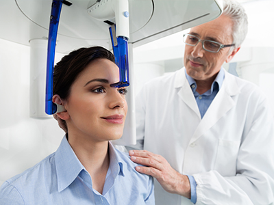
A CBCT appointment is typically quick and straightforward. Patients remain seated or standing while the scanner rotates around their head, capturing the necessary volumetric data in a single pass that usually takes less than a minute. There is no need for uncomfortable bites or lengthy stays in a large scanner—most people find the experience brief and noninvasive.
Because CBCT is targeted, preparation is minimal. Patients may be asked to remove metal objects such as jewelry or removable dental appliances that could interfere with image clarity. Our team provides clear instructions and remains nearby to address questions, ensuring the process is as comfortable and efficient as possible.
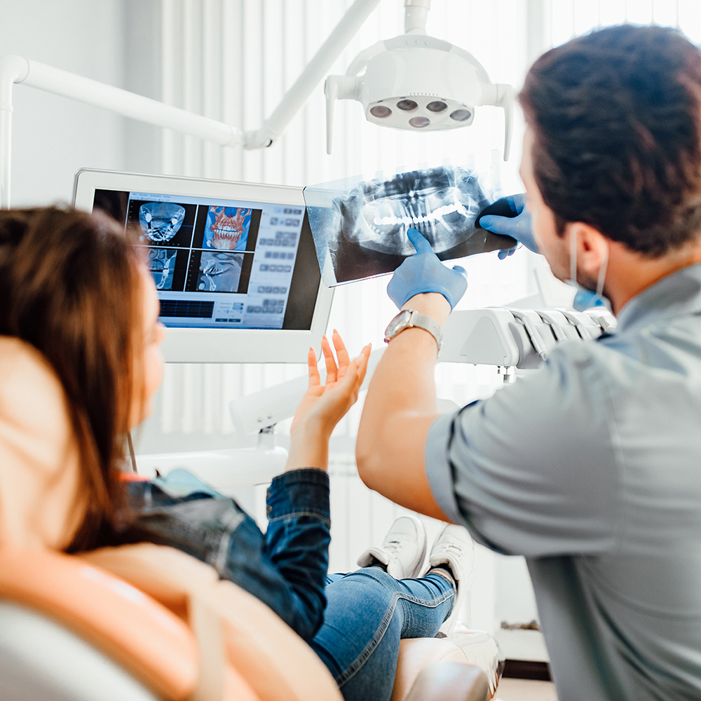
After the scan, the images are reviewed by the treating clinician and incorporated into the treatment plan. We take time to explain findings in patient-friendly terms, show relevant slices or 3D views when helpful, and discuss how the information informs next steps. This collaborative review helps patients understand their condition and participate in decisions about care.
In summary, CBCT is a powerful diagnostic tool that enhances the precision and predictability of modern dental care. When used judiciously, three-dimensional imaging supports safer implant placement, more effective endodontic treatment, and clearer assessment of complex anatomy — all while following dose-conscious practices. If you have questions about how CBCT might be used in your care, please contact us for more information.


Cone-beam computed tomography, commonly called CBCT, is a three-dimensional imaging technique that captures volumetric data of the teeth, jaws, and supporting structures in a single rotation. The scan produces cross-sectional views and 3D reconstructions that reveal spatial relationships between teeth, bone, nerves, and sinuses. Used judiciously, CBCT supplies diagnostic detail that complements conventional two-dimensional radiographs.
At Flossophy, we use CBCT to answer specific clinical questions rather than as a routine scan, which helps target imaging to individual needs. The resulting images support clearer diagnoses and more predictable treatment planning across several dental specialties. Patients benefit from more informed clinical decisions and better communication about proposed care.
Traditional dental X-rays produce two-dimensional images that compress complex anatomy into a flat view, which can obscure overlapping structures and limit depth information. CBCT captures volumetric data so clinicians can view anatomy in axial, sagittal, and coronal planes as well as generate 3D renderings for a complete spatial understanding. This added dimensionality improves detection of features such as root curvatures, fractures, and bone contours that may be difficult to appreciate on 2D films.
CBCT is not intended to replace all conventional radiography; each modality has strengths depending on the diagnostic need. For many routine screenings and bitewing examinations, 2D imaging remains appropriate and lower in radiation. The choice between CBCT and traditional X-rays depends on the question being asked and the level of detail required for safe, effective treatment.
CBCT is commonly recommended when three-dimensional information about bone volume, density, and the location of critical anatomic landmarks is needed for safe implant placement. The scan helps clinicians measure available bone, assess sinus position, and identify the course of the inferior alveolar nerve, all of which guide implant size, angulation, and position. This information reduces intraoperative surprises and supports more predictable restorative outcomes.
In many cases CBCT data are used to design surgical guides that translate virtual implant plans into precise intraoral placement. Guided workflows help align prosthetic goals with surgical execution, improving the likelihood of optimal function and esthetics. The decision to obtain a CBCT is individualized and based on the complexity of the case and the specific planning needs.
CBCT provides detailed visualization of root canal systems, canal curvatures, and periapical anatomy that can be obscured on conventional radiographs. This capability allows clinicians to locate extra canals, detect complex canal morphology, and identify root fractures or resorptive defects more reliably. Better visualization can change treatment approach and increase the chance of successful endodontic therapy.
Three-dimensional imaging is also valuable for assessing the extent of periapical lesions and their relationship to surrounding structures. When indicated, CBCT can help determine whether nonsurgical retreatment, surgical endodontics, or referral to a specialist is the most appropriate next step. As with other indications, CBCT is used selectively to answer targeted clinical questions.
Modern CBCT systems are designed with dose-conscious protocols that balance image quality and radiation exposure. Clinicians select the smallest field of view and the lowest resolution setting that will reliably answer the clinical question, avoiding larger-volume scans when they are not needed. This approach follows the principle of using the lowest radiation dose necessary for diagnosis.
CBCT typically uses lower radiation levels than medical CT for focused head and neck imaging, but any exposure is evaluated against its diagnostic benefit. Our team provides patient education about the scan, positions patients carefully to reduce motion artifacts, and avoids repeat scans whenever possible. These practices contribute to responsible, patient-centered imaging.
A CBCT appointment is generally quick and noninvasive; most scans take less than a minute of actual exposure time while the machine rotates around the head. Patients remain seated or standing and are asked to remove metal objects such as jewelry or removable dental appliances that can cause artifacts. Technicians position patients and provide instructions to stay still to optimize image clarity and reduce the chance of retakes.
After the scan, the treating clinician reviews the images and discusses findings in patient-friendly terms, showing relevant slices or 3D views as needed. The images are incorporated into the treatment plan and used to explain options, anticipated steps, and next actions. If additional specialist input is advisable, the images can be shared to support multidisciplinary care discussions.
CBCT images are first reviewed by the treating clinician who requested the scan and who will integrate the findings into the patient’s care plan. Depending on the complexity of the case, images may also be reviewed or co-interpreted by specialists such as oral and maxillofacial surgeons, endodontists, or ear, nose and throat physicians. Multidisciplinary review helps ensure that all anatomic relationships and treatment implications are considered.
When appropriate, images are exported or shared with colleagues to support coordinated care and consistent planning. Clinicians use the same volumetric dataset as a reference point, which improves communication and reduces ambiguity. Patients are encouraged to ask questions during the review so they understand how the imaging informed the recommended treatment.
CBCT has important diagnostic advantages, but it also has limitations that make it unsuitable for certain indications. The modality has relatively low soft-tissue contrast compared with medical CT and can be affected by metal artifacts from restorations or orthodontic appliances, which may degrade image quality. These factors can limit the usefulness of CBCT for evaluating soft-tissue pathology or in areas with extensive metal that produce scatter.
Certain patient factors, such as pregnancy, require careful consideration and typically prompt clinicians to defer imaging unless it is essential for urgent care. In many routine scenarios, conventional radiographs remain the preferred first step because they answer the clinical question with less radiation. The choice of imaging modality is therefore individualized and based on diagnostic need, image quality expectations, and patient safety.
CBCT provides accurate three-dimensional measurements that are exported to treatment-planning software where virtual implant positions are established relative to anatomic landmarks and prosthetic goals. These virtual plans can be used to design and fabricate surgical guides that translate the planned implant trajectory into the clinical setting. Guides reduce variability and help clinicians place implants in locations that support long-term function and esthetics.
The guided workflow integrates restorative planning with surgical execution, improving communication among the restorative dentist, surgeon, and laboratory team when multiple providers are involved. Using CBCT-derived guides can shorten operative time, increase placement accuracy, and support predictable prosthetic outcomes. As with all tools, careful case selection and planning are essential for optimal results.
Preparation for a CBCT scan is minimal: patients should remove jewelry, eyeglasses, and any removable dental appliances that could interfere with image quality. Wear comfortable clothing without metal fasteners near the head and follow any specific instructions provided by the office regarding recent imaging or medical conditions. If you are pregnant or think you might be pregnant, inform the dental team so the clinician can evaluate the necessity and timing of the scan.
Arrive a few minutes early to allow staff to review your medical history and answer questions about the procedure. During positioning, the technician will provide clear directions to remain still and may use head supports to minimize motion. These simple steps help ensure a single, high-quality acquisition that meets the diagnostic need without repeat exposure.
Ready to schedule your next dental appointment or have questions about our services?
Contacting Flossophy Dental is easy! Our friendly staff is available to assist you with scheduling appointments, answering inquiries about treatment options, and addressing any concerns you may have. Whether you prefer to give us a call, send us an email, or fill out our convenient online contact form, we're here to help. Don't wait to take the first step towards achieving the smile of your dreams – reach out to us today and discover the difference personalized dental care can make.