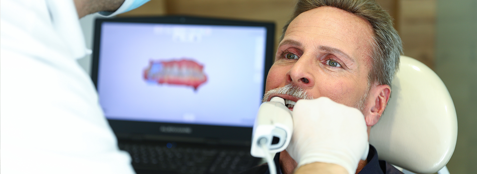
Digital impressions replace the traditional tray-and-putty process with a fast, noninvasive scan of your teeth and surrounding tissues. For many patients, this means avoiding the gagging, taste, or discomfort that can come with conventional impression materials. The scanner gently captures a sequence of images that are stitched together into a highly detailed three-dimensional model, so visits are calmer and more predictable from start to finish.
Beyond comfort, the shift to digital workflows reduces the number of manual steps that once introduced variability. Instead of sending a physical impression through the mail or waiting for a stone model to be poured, clinicians receive an immediate, high-resolution digital file. That streamlines communication between the dental team and the laboratory and reduces opportunities for error caused by handling, distortion, or shipping delays.
This technology is now standard in progressive dental practices because it aligns patient comfort with clinical quality. By prioritizing a gentler chairside experience while delivering precise digital data, practices can keep procedures efficient and patient-focused without sacrificing accuracy.
Intraoral scanners use a handheld wand that emits light and records reflected patterns as it moves across the teeth and soft tissues. Advanced software interprets this optical data in real time, creating a continuous, lifelike 3D representation. The process is non-contact and quick—scans typically take only a few minutes for a full quadrant and a little longer for a complete arch—so patients spend less time in the chair compared with older methods.
As the scanner collects images, the clinician watches a live rendering on-screen, allowing immediate verification of coverage and detail. If a small area needs re-scanning, it can be captured on the spot. This immediate feedback loop helps ensure the final model is complete and clinically useful before the patient leaves the operatory.
Once the scan is finished, the digital file can be refined and annotated using design software. The scanned data preserves essential information—occlusion, margin anatomy, and soft-tissue relationships—that technicians and dentists rely on to design accurate restorations and appliances.
Digital impressions integrate directly with CAD/CAM systems and laboratory software, enabling efficient design and fabrication of crowns, bridges, veneers, and implant restorations. Instead of waiting for a physical model to be manufactured, technicians can begin designing from the digital file immediately. That accelerates collaboration and makes it easier to discuss options or adjustments before any permanent work begins.
The same digital files also support the production of orthodontic and prosthetic appliances—clear aligners, night guards, and surgical guides—using precise, computer-controlled manufacturing methods like milling and 3D printing. The result is a repeatable, predictable process from scan to final product, with fewer manual steps that could introduce fit issues.
For practices that offer in-office milling, digital impressions can lead to same-day ceramic restorations. This capability reduces the number of visits needed for many patients while still maintaining a high standard of precision and esthetics through digitally guided fabrication.
Accurate impressions are the foundation of well-fitting restorations. Even small discrepancies in the data can lead to adjustments, compromised margins, or the need for remakes. Digital scans capture fine anatomical detail in a way that supports precise margin definition and occlusal relationships, helping to produce restorations that seat properly and function comfortably from day one.
A good fit also reduces the risk of secondary problems, such as recurrent decay or gum irritation caused by overhanging or poorly adapted restorations. When restorations and appliances conform closely to the scanned anatomy, patients experience fewer follow-up visits for adjustments and better long-term outcomes for both function and oral health.
Because digital records are stored electronically, clinicians can compare scans over time to monitor wear, shifting, or tissue changes. This longitudinal view supports proactive care—spotting trends early and planning timely interventions that preserve teeth and supporting tissues.
On the day of your appointment, the clinician will explain the process and position the scanner inside the mouth. Breathing, swallowing, and normal tongue movements are fine; the wand is simply moved to capture visible surfaces. Most patients describe the experience as comfortable and unobtrusive. Depending on the scope of treatment, a full-arch digital impression can take just a few minutes, and shorter scans may be needed for crown preparations or targeted restorations.
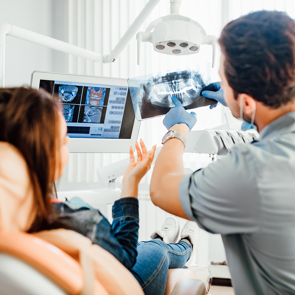
After scanning, the dental team reviews the 3D model with you, pointing out relevant anatomy and discussing next steps. If a restoration or appliance is planned, the team will outline the fabrication pathway—whether that means sending the file to a trusted dental laboratory or using in-office CAD/CAM milling—and what to expect at delivery.
Digital impressions make the treatment pathway more transparent for patients. With visuals on-screen and digital records available for future reference, clinicians and patients can collaborate on decisions with clearer understanding. If you’d like to learn more about how digital impressions are used in restorative or preventive planning, speak with a member of our team during your visit.
Digital impressions combine patient comfort, clinical accuracy, and streamlined communication to support better restorative and appliance outcomes. By capturing high-resolution, three-dimensional data quickly and noninvasively, intraoral scanning reduces manual steps and improves the predictability of treatment workflows from the operatory to the lab.
If you’re curious about how digital impressions could fit into your care plan or want to know what to expect at your next appointment, our practice is ready to explain the technology and walk you through the process. Contact Flossophy Dental to speak with a member of our team and get more information about digital impression services and how they support comfortable, precise dental care.

Digital impressions replace traditional tray-and-putty techniques with a noninvasive optical scan that records the surfaces of teeth and surrounding tissues. A small handheld scanner captures a sequence of images that software stitches into a detailed three-dimensional model. The process is generally faster and more comfortable for patients who find conventional materials unpleasant or gag-inducing.
Because scans produce an immediate digital file, clinicians can review results in real time and confirm coverage before the patient leaves the operatory. The electronic record also simplifies storage and comparison over time, supporting long-term monitoring of teeth and soft tissues. Flossophy's team uses these digital models to enhance communication with dental laboratories and guide precise restorative planning.
Intraoral scanners work by projecting light onto dental surfaces and recording reflected patterns as the wand moves through the mouth. Advanced software interprets this optical data in real time to create a continuous, lifelike 3D rendering on the clinician's screen. Scans are noncontact and typically take only a few minutes for a quadrant and slightly longer for a full arch.
The live rendering allows the clinician to verify coverage and re-scan any missed areas immediately, reducing the need for retakes. Scanned data preserves important details such as occlusion, margin anatomy, and soft-tissue relationships that technicians rely on to design restorations. Post-scan editing and annotation tools further refine the model before design or transfer.
Digital impressions are used for a wide range of treatments including crowns, bridges, veneers, implant restorations, and removable prosthetics. Orthodontic appliances such as clear aligners and surgical guides also begin with an accurate digital scan. Night guards, occlusal splints, and other custom appliances are manufactured from the same high-resolution files.
Because digital files integrate with CAD/CAM and laboratory software, technicians can begin designing restorations immediately after capture. The same digital workflow supports both in-office milling and off-site fabrication using computer-controlled milling or 3D printing. This interoperability helps maintain consistency from the scan to the final appliance or restoration.
Many patients find digital scanning more comfortable than traditional impressions because it eliminates the need for impression trays and putty. The wand is small and noninvasive, and normal breathing and swallowing are usually unaffected during the scan. Shorter scanning times reduce overall chair time and the discomfort associated with prolonged procedures.
Digital impressions are especially helpful for patients with a sensitive gag reflex, young children, or anyone who feels anxious with conventional materials. That said, clinicians will still plan the visit to accommodate individual needs and may use adjunctive techniques to improve access and visibility. Open communication during the appointment helps ensure the fastest, most comfortable scan possible.
Accuracy and precise fit are essential to the longevity and function of restorations, and digital impressions improve both by capturing fine anatomical detail. High-resolution scans provide clear margin definition and accurate occlusal relationships, which reduce the need for extensive chairside adjustments. When restorations seat properly from the start, patients experience better function and fewer follow-up visits.
A well-adapted restoration also lowers the risk of secondary problems like recurrent decay or soft-tissue irritation caused by poor margins. Digital records can be compared across appointments to detect wear, shifting, or tissue changes over time, supporting proactive maintenance. Clinicians use this longitudinal information to plan timely interventions that protect oral health.
Digital impressions can shorten turnaround times because laboratories and design software receive usable files immediately after capture. For practices equipped with in-office milling, the workflow can enable same-day ceramic restorations for appropriate cases. Even when work is sent to an external laboratory, the digital transfer accelerates collaboration and reduces shipping delays.
Faster digital workflows do not eliminate all clinical steps, and the overall timeline still depends on treatment complexity and healing requirements. For multi-stage procedures such as implant care or full-mouth rehabilitation, additional visits remain necessary to ensure proper integration and function. The practice will explain the expected timeline and milestones during treatment planning so patients know what to expect.
Digital impression files are exported in commonly accepted formats and transmitted securely to dental laboratories or kept in-office for CAD/CAM production. Laboratories import the files into their design environments to model restorations, plan implants, or fabricate appliances with precision. In-office systems use the same digital data to drive milling machines or 3D printers for chairside restorations and appliances.
Secure file transfer and proper file management are important to protect patient data and preserve the integrity of the scan. Clinicians and laboratory partners follow established protocols to verify file accuracy and confirm lab instructions before production. Clear communication about margin lines, material choices, and occlusal goals helps reduce revisions and ensures the final product meets clinical requirements.
During a digital impression appointment the clinician will explain the procedure and gently move the scanner throughout the mouth while you breathe and swallow normally. Most full-arch scans take just a few minutes, and targeted scans for a single crown or appliance are even shorter. Patients commonly describe the experience as unobtrusive, and clinicians watch the live model to confirm complete coverage.
After the scan is complete the team reviews the 3D model with the patient, pointing out relevant anatomy and explaining next steps in treatment. If a restoration or appliance is planned, the team will outline whether the file will be sent to a laboratory or used for in-office fabrication. This visual review helps patients understand the plan and participate in decisions about materials and timing.
While intraoral scanning is highly versatile, certain clinical situations may still require traditional impressions or supplemental techniques. Cases with extensive subgingival margins, heavy bleeding, extremely limited mouth opening, or highly reflective surfaces can present scanning challenges. In those scenarios clinicians may use retraction, hemostatic protocols, or combined analog-digital workflows to capture accurate data.
Hybrid approaches are common and allow clinicians to pair the strengths of digital capture with conventional methods when necessary. Choosing the optimal technique depends on the clinical objectives, patient anatomy, and restorative materials involved. Your dental team will recommend the most reliable approach for predictable, high-quality results.
To determine whether digital impressions are appropriate for your care, schedule a consultation so the clinician can evaluate your oral health, restorative needs, and treatment goals. The evaluation typically includes a clinical exam, discussion of previous dental work, and an explanation of how digital scanning would fit into your plan. This collaborative review helps establish expectations and identifies any special steps needed to ensure scan accuracy.
At Flossophy our team will explain the technology, demonstrate a live scan when helpful, and outline the workflow for your specific treatment. We encourage patients to ask questions about what to expect during scanning and how digital records will be used throughout care. If you would like more information about digital impressions during your next visit, ask a member of the clinical team for a demonstration.

Ready to schedule your next dental appointment or have questions about our services?
Contacting Flossophy Dental is easy! Our friendly staff is available to assist you with scheduling appointments, answering inquiries about treatment options, and addressing any concerns you may have. Whether you prefer to give us a call, send us an email, or fill out our convenient online contact form, we're here to help. Don't wait to take the first step towards achieving the smile of your dreams – reach out to us today and discover the difference personalized dental care can make.