Digital radiography is the modern method of capturing dental x-ray images using electronic sensors and computer processing instead of traditional film. For patients, this translates to faster appointments and immediate access to clear images that help clinicians see beneath the surface of a tooth and into surrounding bone. The shift from film to digital is more than a technological update — it changes how quickly clinicians can diagnose problems, plan treatment, and communicate findings with patients in real time.
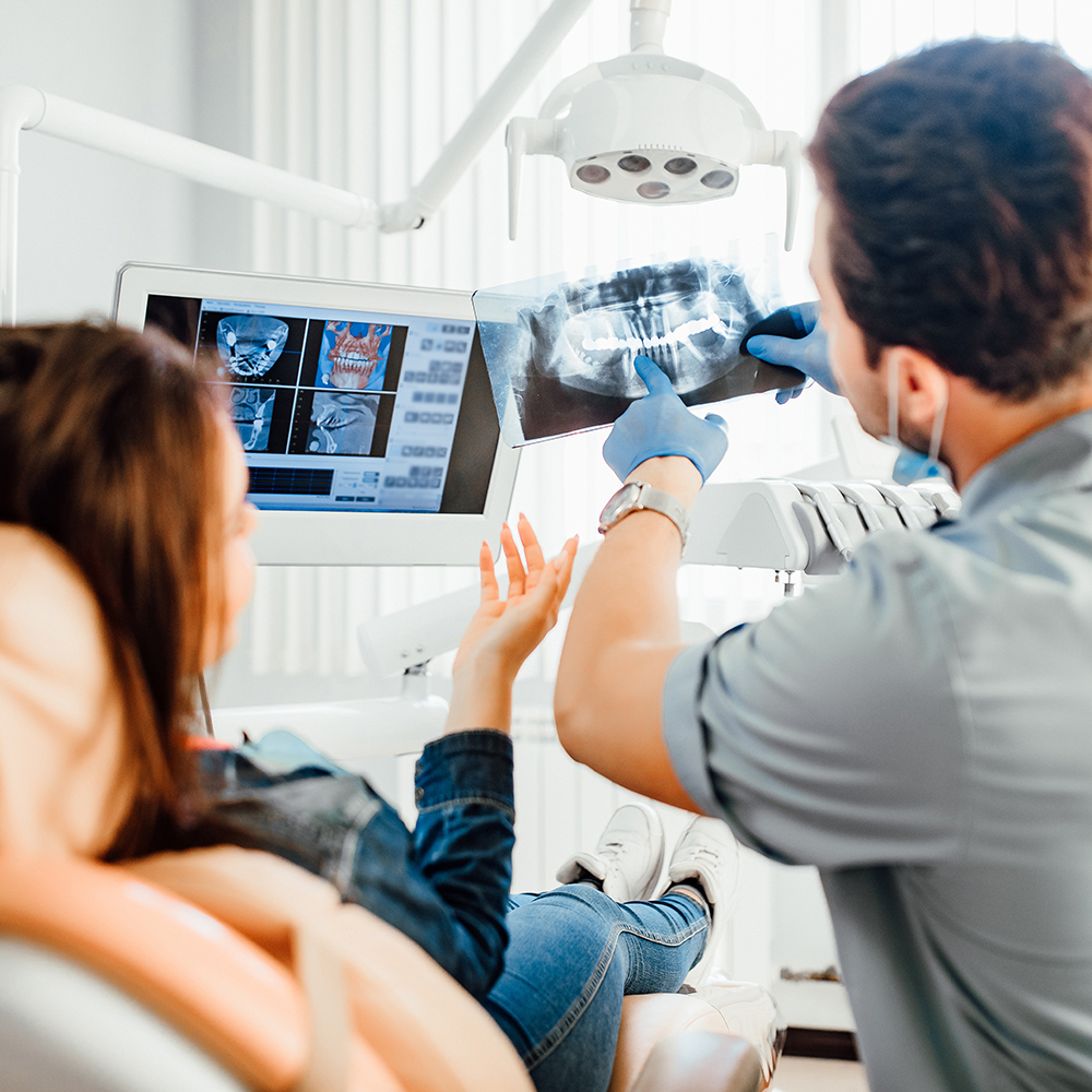
When a clinician obtains a digital radiograph, the image appears on a monitor within seconds, enabling a more conversational approach to care. This instant feedback helps patients understand what the clinician is seeing and why a particular recommendation might be made. Rather than waiting for film to be developed and interpreted later, both patient and provider can review the same image together during the visit, which supports clearer decision-making and peace of mind.
At its core, digital radiography is designed to make dental imaging more efficient and more useful. The images are stored electronically in the patient record, where they can be revisited, compared over time, or forwarded to specialists as needed. Because these files are digital, they integrate seamlessly with other diagnostic tools and clinical software used in modern dental practices.
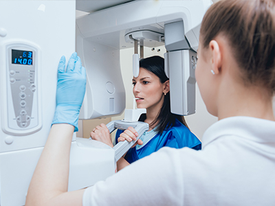
Instead of photographic film, digital radiography uses a flat electronic sensor or a small phosphor plate to detect x-rays. When x-rays pass through the head and teeth, different tissues absorb varying amounts of radiation. The sensor converts these differences into electric signals that are processed by software and rendered as a high-resolution image. This conversion process happens in seconds, delivering a view that can be adjusted for contrast, magnified, or annotated without loss of quality.
There are two common approaches: direct digital sensors that transmit the image instantly to a computer, and semi-direct systems (often called phosphor plate systems) where the plate is scanned after exposure. Both yield diagnostic-quality images with advantages over film, such as adjustable brightness and enhanced detail visibility. Clinicians can use software tools to measure dimensions, detect subtle changes, and store standardized images for future comparison.
Because digital files are adaptable, they can be optimized for different diagnostic tasks. For example, a clinician may apply edge enhancement to highlight carious lesions or adjust brightness and contrast to evaluate bone levels around a tooth. These tools do not replace clinical judgment, but they augment examination by revealing information that might otherwise be harder to detect on conventional film.
One of the most tangible benefits of digital radiography is speed. Images are available immediately, reducing the time a patient spends waiting for results and allowing clinicians to proceed with a treatment plan without delay. This efficiency is especially helpful during restorative procedures, endodontic work, or when monitoring changes over time. Instant imaging supports a more streamlined visit and a better-informed discussion between clinician and patient.
Digital radiography also supports improved collaboration. Images can be shared electronically with specialists, labs, and insurance providers when necessary, facilitating coordinated care without physical film. Multiple clinicians can view the same image concurrently, which is useful for complex cases or when second opinions are requested. The electronic format also simplifies recordkeeping, making it easier to retrieve past images and compare them with current studies.
From a patient perspective, digital images are typically easier to understand. Clinicians can zoom in on areas of concern, annotate findings, and show side-by-side comparisons that illustrate changes over time. This visual clarity helps patients participate in treatment decisions and leaves less room for uncertainty about proposed procedures or the health of their teeth and supporting structures.
Another practical advantage is the reduced need for retakes. Digital sensors often produce clearer images with fewer exposures, so clinicians can reach diagnostic confidence more quickly. While every clinical situation is unique, the improved image quality and on-screen adjustments often decrease the frequency of repeat exposures and can result in a smoother diagnostic workflow.
Digital radiographs are an integral part of comprehensive dental assessment and treatment planning. They provide essential information for routine exams — such as detecting cavities between teeth — and for more involved procedures like implant planning, endodontics, and periodontal assessment. Because digital files can be integrated with other diagnostic modalities, they support a holistic view of oral health that informs long-term care strategies.
For procedures that require precision, such as implant placement, digital imaging can be combined with three-dimensional scans and digital impression data to create a detailed treatment roadmap. Even when three-dimensional imaging is not required, high-quality two-dimensional digital radiographs help clinicians determine the location of roots, the depth of decay, and the status of bone levels, all of which influence the sequence and type of care provided.
Clinicians also use digital images to monitor healing and outcomes. Post-treatment radiographs can verify the success of a root canal, the fit of a restoration, or the stability of bone after a surgical procedure. Because images are stored and organized within the patient record, it is straightforward to compare baseline and follow-up images to assess progress objectively and make informed adjustments to a treatment plan over time.
Digital radiography typically requires lower radiation exposure than traditional film-based methods because sensors are more sensitive and image processing can enhance diagnostic detail without increasing dosage. Clinicians continue to follow the ALARA principle — keeping exposure As Low As Reasonably Achievable — using lead aprons, thyroid collars when appropriate, and modern machines calibrated to minimize dose while producing diagnostically useful images.
Image quality in digital radiography is high and continually improving with better sensors and software algorithms. The ability to manipulate images — changing contrast, magnifying, or applying filters — helps clinicians detect conditions that might be less obvious on conventional film. These capabilities refine diagnosis and support clinical decisions without subjecting patients to extra exposures solely for better visualization.
There are also environmental benefits to consider. Digital systems eliminate the need for chemical developers and film disposal, reducing hazardous waste associated with traditional radiography. The electronic format reduces physical storage needs and simplifies records management, which can be both environmentally and operationally advantageous for a busy practice.
Taken together, the safety profile, diagnostic utility, and practical benefits of digital radiography have made it a standard component of contemporary dental care. By combining reliable imaging technology with clinical expertise, dental teams can deliver clearer diagnoses and more predictable treatment outcomes while minimizing environmental impact.
In summary, digital radiography brings faster, clearer, and more adaptable imaging to everyday dental care. It supports efficient clinical workflows, enhances communication between clinician and patient, and integrates smoothly into comprehensive treatment planning. If you would like to learn more about how digital x-rays are used in our office or how they support specific treatments, please contact us for more information.


Digital radiography is the modern method of capturing dental x-ray images using electronic sensors and computer processing instead of traditional film. It uses flat sensors or phosphor plates to record the pattern of x-rays that pass through teeth and supporting structures. Software converts those signals into high-resolution images that appear on a computer screen within seconds.
Because images are available immediately, clinicians can review findings with patients in real time and make faster diagnostic decisions. Digital files are stored in the patient record, where they can be compared over time or sent to a specialist if needed. The technology has become a standard tool in contemporary dental care for routine exams and advanced procedures.
When x-rays are taken, sensors detect differences in radiation absorption across tissues and convert those differences into electrical signals. Direct sensors send the data instantly to a computer, while phosphor plate systems store the latent image that is later scanned and digitized. Advanced software processes these signals into images that can be adjusted for contrast, magnification, and measurement.
Clinicians can apply filters and measurement tools to enhance the visibility of caries, root anatomy, and bone levels without degrading image quality. These adjustments help reveal subtle changes that might be harder to see on film, but they do not replace clinical judgment. The speed and flexibility of digital processing make it easier to capture diagnostically useful views with fewer repeat exposures.
Patients benefit from digital radiography through shorter appointment times and quicker diagnosis because images appear immediately. The improved image quality and on-screen tools make it easier for clinicians to explain conditions and for patients to visualize treatment needs. Digital systems also tend to reduce the need for retakes by producing clearer images on the first attempt.
Electronic images support better communication with specialists and labs, enabling coordinated care when procedures require outside consultation. Side-by-side comparisons and annotation tools help patients follow changes over time and participate in treatment decisions. Overall, the combination of speed, clarity, and shareability enhances the patient experience.
Digital radiography generally requires less radiation than traditional film because modern sensors are more sensitive and software can enhance image detail. Dental teams follow the ALARA principle—keeping exposure As Low As Reasonably Achievable—by using appropriate settings and protective measures such as lead aprons and thyroid collars when indicated. Machines are calibrated and maintained to produce diagnostically useful images at the lowest reasonable dose.
Because image processing can improve diagnostic visibility, clinicians often avoid increasing dose to obtain clearer images and instead rely on software adjustments. Repeat exposures are less common with digital sensors, which further reduces cumulative exposure over time. X-rays are prescribed only when clinically necessary as part of a comprehensive exam or treatment plan.
There are two common sensor approaches in digital radiography: direct digital sensors (CCD/CMOS) and phosphor plate systems (PSP). Direct sensors transmit images instantly to the computer, offering immediate feedback, while phosphor plates are scanned after exposure to produce the digital image. Each system provides diagnostic-quality images but differs in workflow, comfort, and initial hardware requirements.
Clinicians select the sensor type based on the clinical need, patient comfort, and the practice’s existing equipment and software. Both systems allow image enhancement and integration with electronic records, so the choice rarely limits diagnostic capability. Ongoing advances in sensor design continue to narrow any performance differences between platforms.
Digital radiographs improve diagnosis by revealing interproximal decay, root anatomy, and bone levels with clarity and the ability to magnify areas of interest. High-resolution images assist in detecting early changes that might not be visible on a clinical exam alone, which helps prioritize treatment. This information is essential for procedures like root canals, periodontal assessment, and restorative planning.
For implant planning and other precision procedures, two-dimensional digital images are often combined with three-dimensional scans and digital impressions to create a comprehensive treatment roadmap. Post-treatment radiographs provide objective data for evaluating healing and restoration fit over time. The ability to archive and compare images supports evidence-based adjustments to care plans.
Digital radiographs can be shared electronically with specialists, laboratories, and referring providers to support coordinated care and second opinions. Standard file formats and secure transfer protocols make it straightforward to send images without physical film. Electronic sharing reduces delays in communication and facilitates collaborative treatment planning.
When patients request copies of their images, offices typically provide electronic files or printed versions as appropriate for the recipient. Transfers are completed with attention to privacy and file integrity so the receiving clinician has diagnostically useful data. If a specialist needs a high-resolution copy for planning, digital files can be exported in formats compatible with most clinical imaging systems.
Digital radiographs are stored in the patient’s electronic record, where they are organized, indexed, and retained for clinical use and comparison over time. Modern dental record systems include access controls, audit trails, and encryption to protect patient information in accordance with privacy regulations. Regular backups and secure hosting reduce the risk of data loss and ensure long-term availability of images.
Patients may request access to their radiographs and clinicians can provide copies for referrals or personal records, subject to privacy safeguards. Retained images create a reliable baseline for monitoring changes and assessing treatment outcomes across multiple visits. Proper recordkeeping improves continuity of care and supports informed clinical decisions.
Preparing for a dental x-ray generally requires little to no special preparation from patients beyond standard hygiene and removing jewelry in the head and neck area. Pregnant patients should inform the dental team so clinicians can evaluate the necessity and timing of radiographs and apply extra protective measures when appropriate. Children are positioned carefully and may require additional assistance to obtain the correct view while minimizing exposure.
The dental team will explain the process, position the sensor or plate, and use protective equipment as needed before taking any images. Images are reviewed immediately to confirm diagnostic quality so that retakes are minimized and the visit can proceed efficiently. If you have concerns or questions about the procedure, the staff will address them before imaging begins.
At Flossophy, digital radiography is integrated into routine exams and specialty procedures to enhance diagnostic accuracy and patient communication. Clinicians use on-screen images to explain findings during the visit and to plan treatments such as restorations, root canals, and implant placement. The practice combines radiographs with other digital tools to create coordinated, patient-centered care.
Images are stored in the electronic patient record for follow-up comparison and can be forwarded to specialists when collaborative care is needed. If you prefer to review your images in detail, the team at our Bourbonnais office at 750 Almar Pkwy, Suite 101 will take time to walk through them and answer questions. Contact the office if you would like to learn more about how digital imaging is used in your care.
Ready to schedule your next dental appointment or have questions about our services?
Contacting Flossophy Dental is easy! Our friendly staff is available to assist you with scheduling appointments, answering inquiries about treatment options, and addressing any concerns you may have. Whether you prefer to give us a call, send us an email, or fill out our convenient online contact form, we're here to help. Don't wait to take the first step towards achieving the smile of your dreams – reach out to us today and discover the difference personalized dental care can make.