An intraoral camera is a compact, pen-sized imaging device designed specifically for the oral environment. It captures high-resolution, full-color images of teeth, gums, restorations and other soft tissues inside the mouth. Because the camera sits close to the surface it records, it can reveal subtle details—hairline cracks, early enamel wear, small areas of decay, worn margins around fillings, and inflammation of the gums—that are difficult to see with the naked eye alone.
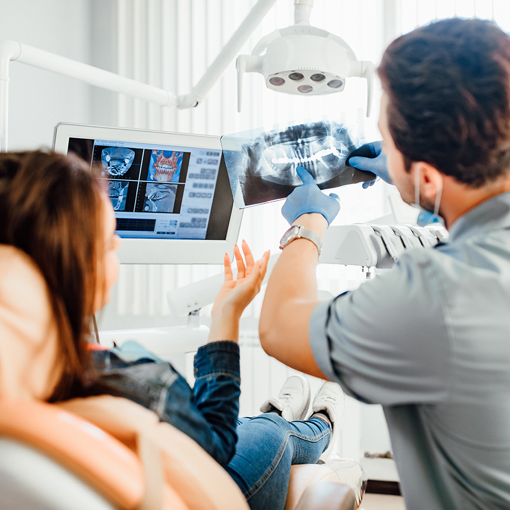
Those clear, magnified images help both clinicians and patients understand what’s happening inside the mouth in real time. Rather than relying solely on verbal descriptions or feeling uncertain about a diagnosis, a patient can view the same images the clinician sees and follow along as findings are explained. The visual nature of the information improves comprehension and supports more informed decision-making about recommended care.
Because intraoral cameras produce true-color photographs, they also provide context that black-and-white radiographs do not: surface texture, oral mucosa appearance, plaque accumulation and the condition of existing dental work are all visible. When used alongside other diagnostic tools, intraoral images make it easier to form a complete, accurate picture of oral health.
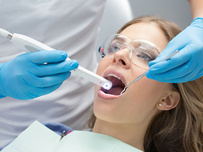
During a routine exam or a focused evaluation, the clinician gently guides the intraoral camera into the mouth to photograph specific areas of interest. The device connects to a chairside monitor so images appear immediately and can be enlarged, compared side-by-side, or annotated for clarity. Operation is fast and noninvasive: most image captures take only a few seconds and are entirely comfortable for the patient.
Images can be frozen, reviewed frame-by-frame, and magnified without losing clarity, which helps the team point out exact areas of concern. If a clinician wants to show how a problem looks from different angles, they can capture multiple views during the same visit. This live visual feedback supports focused conversations and helps patients see why a particular recommendation may be appropriate.
Because the camera is small and maneuverable, it works well for both adult and pediatric exams. It also integrates smoothly with other digital systems in the office, allowing the clinician to pull past images for comparison, overlay notes, and document the mouth’s condition at each visit. The process is streamlined so diagnostic imaging adds value without significantly extending appointment time.
Intraoral imaging enhances diagnostic accuracy by providing a close-up, detailed record of oral conditions. Early lesions and marginal breakdowns that may be missed in a quick visual exam are often visible with magnification, which supports earlier, more conservative interventions. When problems are identified earlier, treatment can be planned with an emphasis on preserving natural tooth structure and maintaining long-term function.
For treatment planning, these images are invaluable. They help the clinical team explain the rationale for proposed procedures, show treatment sequencing, and illustrate expected outcomes. When multiple treatment options exist, visual documentation can make it easier for patients to weigh choices and understand trade-offs. The clarity and immediacy of intraoral images also facilitate coordination with specialists or laboratory partners when complex restorative or surgical work is needed.
Additionally, intraoral cameras support quality assurance. Before, during and after treatment, photographs document the condition of restorations and soft tissues, which creates a clear record of the care provided and its outcomes. This photographic documentation reinforces clinical decisions and helps the team track healing and the longevity of interventions over time.
Images captured with an intraoral camera are saved directly into the patient’s electronic record so they are available for future appointments and long-term monitoring. Stored images can be labeled, dated and grouped by area, allowing clinicians to quickly review historical changes and trends. This chronological record is especially useful for tracking the progression or resolution of conditions over successive visits.
Because saved images are high-resolution and standardized, they can be shared with other dental professionals when collaborative care is needed. Whether referring to a specialist, consulting a dental laboratory about a restoration, or documenting a treatment sequence, the photographic record provides an objective visual reference. That helps ensure continuity and accuracy when multiple providers are involved in care.
For administrative and clinical clarity, these images also support documentation of findings discussed during appointments. Clear photographic records reduce ambiguity in clinical notes, making it easier for the team to communicate internally and to maintain consistent, thorough care for each patient over time.
When an intraoral camera is used during your visit, expect a more interactive examination. Our team can pause to show you images on the monitor, point out specific findings, and explain how various issues might be addressed. This visual approach fosters a collaborative conversation in which the clinician’s observations and the patient’s questions are anchored to the same images.
Using intraoral photography also improves transparency in care. Because you can see problems directly, treatment recommendations are easier to understand and follow. Likewise, images taken over time demonstrate how conditions respond to home care, preventive measures, or clinical treatment, which helps build a practical, evidence-based plan for maintaining oral health.
At Flossophy Dental, we incorporate intraoral imaging into exams and consultations as appropriate to enhance diagnostics and patient education. Our goal is to combine modern imaging tools with clear communication so every patient feels well-informed about their oral health and confident in the path forward.
In summary, intraoral cameras are powerful diagnostic and communication tools that bring clarity to dental examinations. They reveal fine detail, support precise treatment planning, create enduring clinical records, and make appointments more collaborative. For more information about how intraoral imaging may be used during your next visit, please contact us.


An intraoral camera is a small, pen-sized digital imaging device designed to photograph the inside of the mouth. It captures high-resolution, full-color images of teeth, gums and restorations from close range. Clinicians use it to document surface texture and subtle changes that may be difficult to see with the naked eye.
Unlike traditional black-and-white radiographs, intraoral images show true color and surface detail, including plaque, mucosal changes and the condition of fillings. The camera complements radiographs and visual exams rather than replacing them. Together, these tools create a more complete diagnostic picture for accurate assessment.
An intraoral camera can reveal hairline cracks, early enamel wear, small areas of decay and deteriorating margins around restorations. It also makes soft tissue findings such as inflammation, lesions and plaque accumulation more visible. These subtle signs are often hard to document during a quick visual exam but are obvious when magnified and photographed.
Because the camera records true-color images, clinicians can evaluate surface texture and mucosal appearance in addition to hard-tissue issues. This broader visual context helps differentiate between cosmetic concerns, minor changes and findings that require clinical intervention. Early detection through imaging often enables more conservative treatment options.
During an exam the clinician gently guides the intraoral camera into the mouth to photograph areas of interest while images display on a chairside monitor. Image capture is fast and noninvasive, and most photographs take only a few seconds to record. The live feed lets the clinician move the device to capture multiple angles as needed for a thorough view.
Captured images can be frozen, enlarged and annotated so specific findings are easy to point out and explain. Multiple views can be compared side-by-side to illustrate change or to show how a problem appears from different perspectives. This streamlined workflow adds diagnostic value without significantly extending appointment time.
Intraoral cameras are designed for comfort and are well tolerated by both adults and pediatric patients. The device is small, lightweight and used briefly to capture images, so most patients experience no discomfort during imaging. Standard infection-control barriers and protocols are used to ensure safe operation.
The technique involves no radiation, and it is entirely noninvasive, making it an excellent adjunct for routine exams and focused evaluations. Because captures are quick, intraoral imaging typically does not add meaningful time to an appointment. Clinicians can adjust positioning and pace to accommodate anxious or very young patients.
High-resolution intraoral photographs enhance diagnostic accuracy by revealing fine detail such as early lesions and marginal breakdowns that might be missed during a cursory exam. Identifying problems earlier supports more conservative treatment choices aimed at preserving tooth structure and function. Photographic documentation also helps clinicians monitor progression or healing over time.
For treatment planning, images make it easier to explain the rationale for proposed procedures and to illustrate sequencing and expected outcomes. Visual documentation also facilitates coordination with specialists and dental laboratories when restorative or surgical work is needed. Clear images support consistent, evidence-based decisions throughout care.
Yes. Images captured with an intraoral camera are typically stored directly in the patient's electronic record so they remain available for future appointments and long-term monitoring. Stored images can be labeled, dated and grouped by area to document changes and trends over time. This chronological record is especially useful for tracking the progression or resolution of conditions.
Because the photographs are standardized and high-resolution, clinicians can use them to compare past and present findings quickly and objectively. The photographic record also reduces ambiguity in clinical notes and supports clear internal communication among the care team. When needed, these images can be referenced for follow-up care or treatment evaluation.
Intraoral imaging creates a shared visual reference that helps patients and clinicians see the same findings at the same time. This live, point-by-point view anchors conversations and makes explanations more concrete than verbal descriptions alone. As a result, patients tend to leave the appointment with a clearer understanding of their oral health status.
The visual approach supports shared decision-making by illustrating trade-offs and expected outcomes for different treatment options. It also streamlines informed-consent discussions because the images document the issues being addressed. Overall, imaging improves transparency and helps build confidence in the recommended plan of care.
Yes. Because intraoral photographs are high-resolution and standardized, they can be exported or shared with other dental professionals when collaborative care is needed. Secure sharing of images helps ensure that specialists receive accurate visual information before a consultation or procedure. Photographs can also accompany laboratory prescriptions to improve communication about shade, margin condition and preparation details.
Using consistent photographic records reduces the risk of miscommunication and helps specialists and lab technicians plan restorations or procedures more precisely. When referring care, clinicians often include annotated images and notes to provide context and streamline coordination. This practice supports continuity and accuracy across multi-provider treatment plans.
Intraoral cameras help clinicians detect early signs of disease, such as small carious lesions, enamel wear and plaque accumulation, which supports timely preventive measures. By making these issues visible, imaging reinforces personalized home-care recommendations and motivates patients to address problem areas. Regular photographic comparisons can highlight improvements or areas that need additional attention.
Because images document the mouth's condition at each visit, clinicians can tailor recall intervals and preventive strategies based on objective evidence. Imaging also aids in monitoring mucosal changes and oral hygiene practices over time. This proactive approach increases the likelihood that minor issues will be managed before they require more extensive intervention.
Expect a more interactive, educational exam in which the clinician may pause to show you real-time images on a monitor and explain specific findings. The process is quick and noninvasive: the camera is guided into the mouth to capture targeted views while you watch the images as they appear. Clinicians often annotate or enlarge captured frames to point out exact areas of concern and to discuss recommended next steps.
Images are saved to the patient's record and can be reviewed at subsequent visits to chart progress or healing. At our Bourbonnais, IL office, photographic documentation is used to support clear communication, continuity of care and informed decision-making. If you have questions about images taken during your visit, the clinical team will review them with you and explain how they inform your personalized care plan.
Ready to schedule your next dental appointment or have questions about our services?
Contacting Flossophy Dental is easy! Our friendly staff is available to assist you with scheduling appointments, answering inquiries about treatment options, and addressing any concerns you may have. Whether you prefer to give us a call, send us an email, or fill out our convenient online contact form, we're here to help. Don't wait to take the first step towards achieving the smile of your dreams – reach out to us today and discover the difference personalized dental care can make.